Myosis is a pupil constriction reflex that reduces retinal illumination and can alter the balance of ocular muscles. In people with Strabismus, commonly called crossed eyes, the misalignment is already fragile, so the added effect of myosis often worsens binocular coordination.
Why Myosis Matters for Eye Alignment
When the pupil narrows, the iris sphincter pulls inward, slightly changing the eye’s rotational axis. This shift is usually negligible, but in a system where the extraocular muscles are already fighting for dominance, even a few degrees of rotation can tip the scale. The four rectus muscles and two oblique muscles coordinate to keep eyes on the same target; myosis can change the pull on the medial and lateral rectus, making an eye drift inward or outward.
Interaction Between Myosis and Strabismus
Strabismus itself is categorized by the direction of deviation: esotropia (inward), exotropia (outward), hypertropia (upward), and hypotropia (downward). Myosis tends to accentuate esotropia because the constricted pupil pulls the eye slightly toward the nose. For patients with exotropia, the effect can be less dramatic but still disrupt binocular vision, the brain’s ability to fuse two images into one.
Symptoms to Watch For
- Increased double vision (diplopia) when moving from bright to dim environments.
- Head tilt or turn to compensate for the misalignment.
- Eye strain and fatigue after reading or using screens.
- Reduced depth perception, especially in low‑light settings.
Because myosis is a reflex to light, these symptoms often flare up at dusk, in classrooms, or when driving at night.
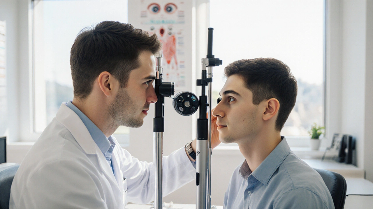
How Professionals Diagnose the Combined Issue
An ophthalmologist or pediatric eye specialist will perform a series of tests:
- Cover‑uncover and alternate‑cover tests to gauge the magnitude of deviation.
- Pupil size measurement under varying luminance to quantify myosis.
- Accommodative‑convergence testing, since accommodation often drives pupil constriction.
- Retinal imaging to rule out optic nerve or retinal disease that could mimic symptoms.
Treatment Options - What Works Best?
The goal is to stabilize both the pupil size and the eye’s alignment. Below is a quick look at the most common approaches.
| Option | Primary Benefit | Typical Duration | Potential Drawbacks |
|---|---|---|---|
| Vision Therapy | Improves binocular coordination and reduces reliance on pupillary reflex. | 12-24 weeks, 1‑hour sessions weekly. | Requires patient commitment; progress can be slow. |
| Prism Glasses | Instantly realigns images, easing diplopia. | Immediate; lenses may be adjusted quarterly. | Can cause peripheral distortion; not a permanent fix. |
| Strabismus Surgery | Corrects muscle imbalances surgically. | One‑time procedure; recovery 4-6 weeks. | Risk of over‑ or under‑correction; possible need for repeat. |
| Pharmacologic Agents | Modulates pupil size (e.g., pilocarpine) to reduce excessive myosis. | Variable; often used as adjunct. | Side effects include blurred near vision and headache. |
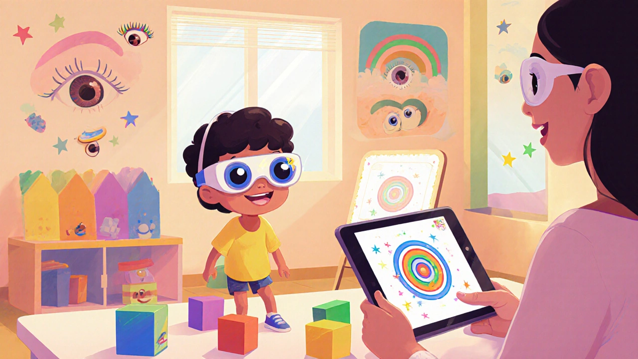
Vision Therapy: The Core Rehabilitation Method
Therapy focuses on exercises that train the brain to ignore the reflex‑driven pupil change and re‑establish stable accommodation‑convergence coupling. Typical drills include:
- Near‑far jump cards to improve rapid focus shift.
- Barrel card or stereogram work for depth perception.
- Computer‑based gaze tracking that flags excessive pupil constriction.
Research from the American Academy of Ophthalmology (2023) shows a 68% success rate when therapy is combined with low‑dose pilocarpine to control myosis.
Prism Glasses: Quick Symptom Relief
Prisms are ground into spectacle lenses, shifting the image toward the affected eye. They don’t address the underlying muscle imbalance but can be lifesavers for night‑time drivers or students who need immediate clarity. An experienced optometrist determines the diopter strength based on the measured deviation in prism diopters.
Surgical Intervention: When Alignment Needs a Fix
When non‑invasive methods fail, surgeons adjust the insertion points of the extraocular muscles. The procedure directly counters the myosis‑induced rotation by tightening or loosening specific muscles. Post‑op, patients often still need vision therapy to cement the new alignment.
Everyday Strategies to Co‑Exist with Myosis and Strabismus
- Use adequate ambient lighting; bright desk lamps reduce pupil constriction spikes.
- Take regular screen breaks (20‑20‑20 rule) to lower eye strain.
- Wear anti‑glare coatings on glasses to minimise sudden light changes.
- Practice head‑position awareness - a subtle turn can compensate for misalignment without fatigue.
Related Concepts and Where to Go Next
The interplay between pupil dynamics and eye alignment also appears in conditions like amblyopia (lazy eye) and nystagmus. Understanding these links helps clinicians choose the right combination of therapy and corrective lenses. If you’re curious about why some children develop crossed eyes after a concussion, look into the broader topic of neurological causes of strabismus. Future posts will dive deeper into surgical techniques and emerging pharmacologic agents.
Key Takeaways
Myosis can tip the delicate balance of eye muscles in people with strabismus, leading to worse diplopia and depth‑perception issues. Early detection, a mix of vision therapy, prisms, and, where needed, surgery, offers the best chance for stable binocular vision. Simple lifestyle tweaks further reduce the burden.
Frequently Asked Questions
Can myosis cause strabismus, or only worsen it?
Myosis alone rarely creates a full‑blown strabismus, but its effect on the iris can shift eye rotation enough to aggravate an existing misalignment. In children with a predisposition, chronic pupil constriction can accelerate the development of crossed eyes.
Is it safe to use eye drops that dilate the pupil for strabismus patients?
Dilating drops can temporarily reduce the myotic pull on the eye, but they may also blur near vision and cause photophobia. They are usually reserved for diagnostic purposes rather than long‑term management.
How long does vision therapy typically take to show improvement?
Most patients notice measurable gains after 8‑12 weeks of consistent weekly sessions, though full stabilization may require 6‑12 months, especially when myosis is severe.
Are prism glasses covered by insurance in Australia?
Many private health funds provide partial reimbursement for therapeutic prisms when prescribed by an ophthalmologist or orthoptist. Public hospital schemes may cover them if the patient meets specific criteria.
What are the signs that surgery might be necessary?
If the eye deviation exceeds 30 prism diopters, if diplopia persists despite therapy and prisms, or if the misalignment interferes with daily activities (e.g., driving at night), an ophthalmic surgeon will likely recommend corrective surgery.


Jay Ram
27 September / 2025I've seen a few patients where the constricted pupil really throws off the muscle balance, making the esotropia jump a degree or two. It can be subtle, but over a day it adds up, especially in low‑light rooms. Keeping the lighting steady helps a lot, and a quick check with a handheld pupil meter can catch the shift early.